
Introducing the L|neMic 1K Ultra!
Our first complete microscope, designed for ultrafast 3D imaging of relatively large objects, such as organoids or spheroids. The L|neMic 1K Ultra allows confocal imaging at frame rates of up to 1 kiloHertz (yes, 1000 high-quality confocal images per second). This unparalleled imaging speed is achieved by focusing the laser beam into a line, rather than into a single spot, increasing the imaging rate up to 1000x in comparison with standard laser-scanning confocal microscopes. Contact us at info@innovativebioimaging.com for technical and pricing information. A scientific article describing the imaging principle is being readied for publication (https://www.biorxiv.org/content/10.1101/2023.04.11.536163).
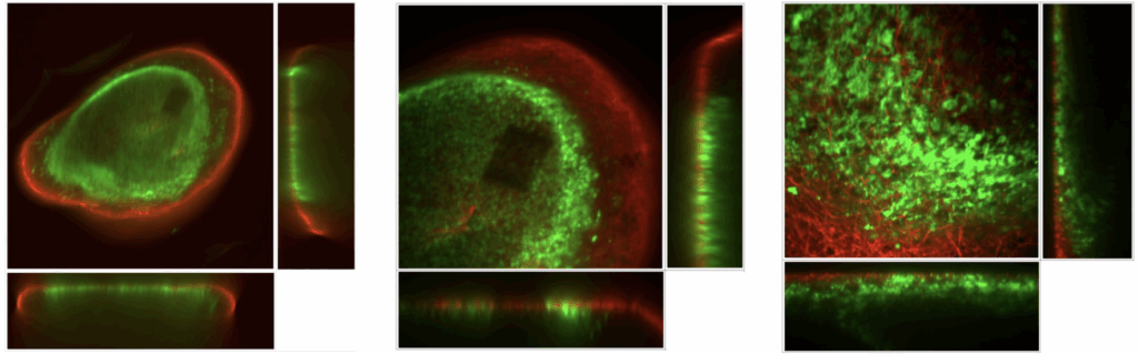
3D reconstructions of a fluorescently labeled organoid imaged by the L|neMic 1K Ultra at three different resolutions (using a 10x, 25x and a 60x objective lens). Individual images took 20 milliseconds to acquire, allowing acquisition of a full z-stack in approximately 1 second. Even faster acquisitions are possible and, in fact, easy.
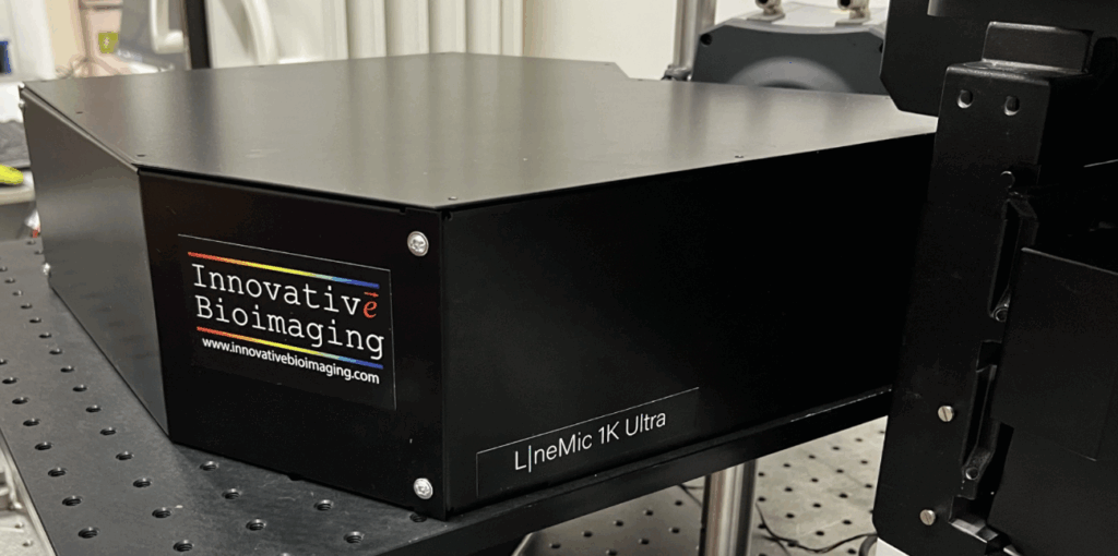
The L|neMic 1K Ultra can be controlled by microManager, but also by the Olympus/Evident CellSens software, providing the user a choice between a free, flexible software environment or a more polished commercial software solution. Up to four excitation wavelengths can be provided. The L|neMic 1K Ultra can perform automated imaging in the 8-, 24-, 96- and 384-well format. For more technical and pricing information, contact info@innovativebioimaging.com.
Coming soon: FL!Ps™
FL!Ps are molecular probes developed specifically for polarization microscopy. The probes allow easy, quantitative imaging of many molecular processes of cell signaling (activation/inactivation of GPCRs, G proteins, GTPases, receptor tyrosine kinases,…)
Check out our preprint:
Miclea P. & al., https://www.biorxiv.org/content/10.1101/2024.02.23.581811
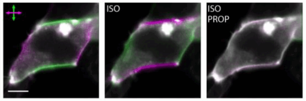
From the preprint:
Imaging activation/inactivation of the beta2-adrenergic receptor, using a FL!P biosensor and a confocal microscope equipped with our 1P FL!Pper module:
Coming soon: DynaMic
A first commercial microscope dedicated to imaging FL!P biosensors, as well as other types of biosensors. The DynaMic, currently under development, will allow fast, sensitive imaging of all the molecular processes observable by FL!Ps, and many more! Keep checking for more information!
1P FL!Pper™
Our rapid polarization modulator rotates the excitation beam polarization between acquisition of individual lines (or even pixels) by a laser scanning confocal microscope. This way, each pixel is imaged twice, using two different polarizations, within one image. Because information with both polarizations is acquired virtually simultaneously, there are no problems caused by cell movement, and even rapid molecular events can be reliably observed and monitored. The 1P FL!Pper is compatible with the full range of visible excitation wavelengths (405-640 nm). In combination with our accompanying Polaris+ software, the 1P FL!Pper it allows easy, accurate quantification of linear dichroism. To learn more about the 1P FL!Pper, including pricing information, contact our sales team.
2P FL!Pper™
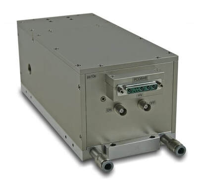
Similar to our 1P FL!PPer, but tailored for two-photon excitation laser wavelengths (700-1200 nm). Unlike single-photon excitation, two-photon excitation allows not just observations of linear dichroism, but accurate determinations of orientations of fluorescent molecules with respect to membranes or filaments. The 2P FL!Pper is compatible with virtually any existing two-photon microscope. The accompanying Polaris+ software allows not only processing of acquired images and display of linear dichroism as color hues on various scales and in various color combinations, but also quantitative measurements of linear dichroism in living cells and determinations of molecular orientations. To learn more about the 2P FL!Pper, including pricing information, contact our sales team.

Polaris/Polaris+ software
Our polarization modulators come with software that allows facile display and quantitation of linear dichroism: Polaris (for analyzing any pair of images acquired with distinct excitation light polarizations) and Polaris+ (for analyzing images acquired with our 1P FL!Pper or 2P FL!Pper polarization modulators). To learn more about the Polaris/Polaris+, including pricing information, contact our sales team.
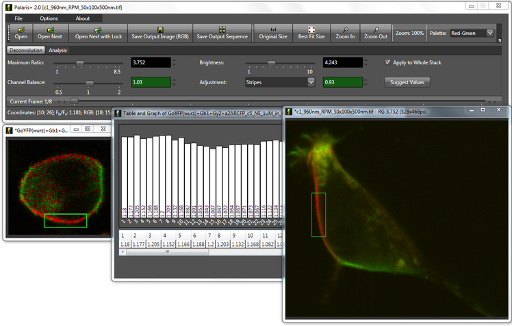
Both versions provide sophisticated, yet easy to use tools that will allow even subtle linear dichroism to be reliably observed and measured. Our software builds on years of expertise in polarization microscopy, and will provide you with both published and proprietary scientifically correct algorithms for image processing and analysis. Both versions of our software allow a wide range of choices for visualizing linear dichroism, including choices suitable for people with color vision impairments. Polaris+ has been designed to streamline processing of time series and z-stacks. Polaris+ takes advantage of our detailed knowledge of all parts of a polarization microscope to deliver results that cannot be matched.
Thanks to our software, you can have a complete polarization microscopy system, capable of delivering publication-quality results, right after installation.